Lung surgery built around the breaths you'll take for the rest of your life — keyhole first.
VATS keyhole technique removes tumours through small incisions, protecting lung function and speeding recovery. Open surgery is chosen when it genuinely serves you — never by default.
What is thoracic oncology?
This region covers cancers of the lung, the mediastinum (the space between the lungs, home to the thymus), the pleura (the lining of the lung and chest cavity, where mesothelioma arises) and the chest wall.
The philosophy here is keyhole-first. VATS — video-assisted thoracoscopic surgery — achieves the same cancer operation as open surgery through small cuts: less pain, faster mobilisation, quicker recovery. When anatomy or stage demands an open approach, that's stated plainly, with reasons.
And because many people facing this region are current or former smokers, support with quitting is woven into care — offered without judgment, always.
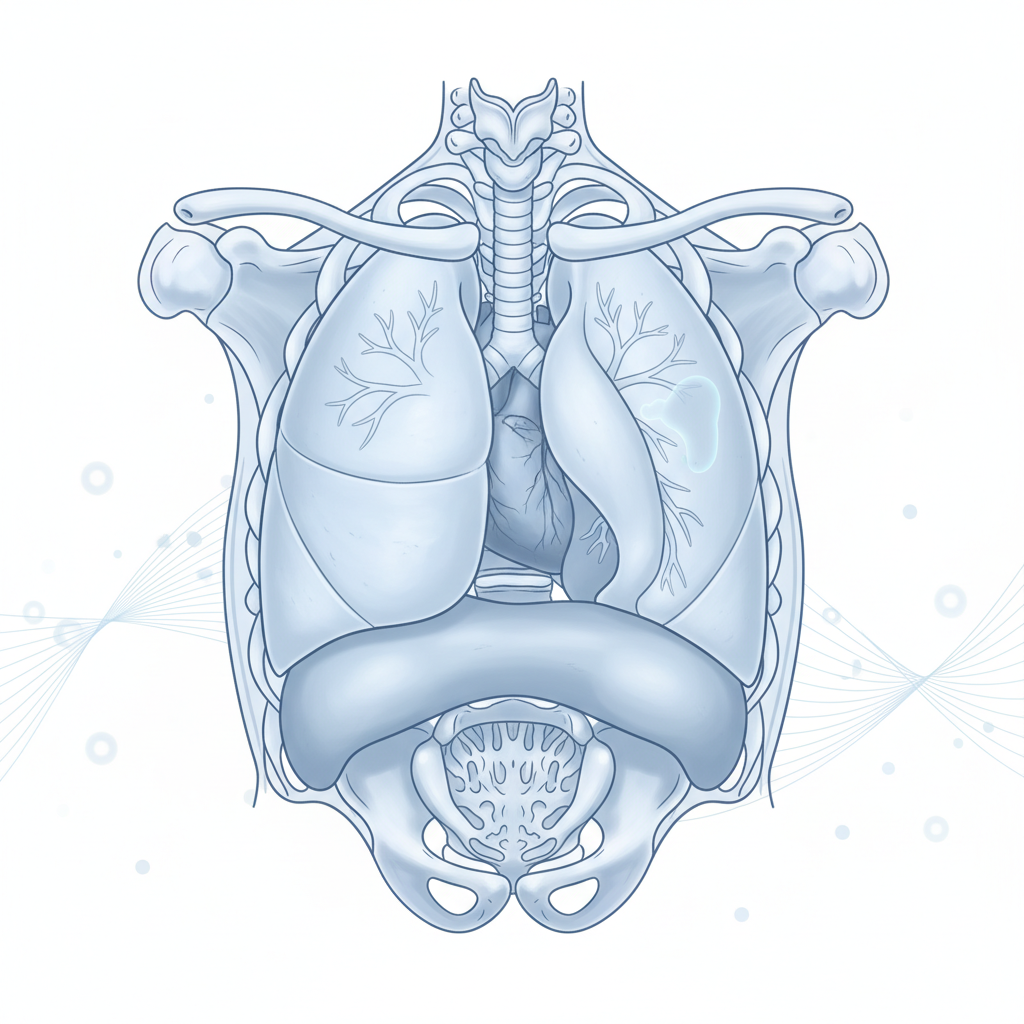
Eight signals from the chest
None of these mean cancer on their own — but each deserves a proper look rather than another round of waiting.
- ①A cough lasting more than three weeks that simply will not settle.
- ②A change in breathlessness — stairs or slopes suddenly harder than before.
- ③Blood-streaked sputum, even once.
- ④Chest pain on one side that persists.
- ⑤Hoarseness without a cold to explain it.
- ⑥Pneumonia recurring on the same side — treated twice at the same spot.
- ⑦Finger clubbing — nails curving gently over rounded fingertips.
- ⑧A long smoking history alongside any of the above — mention it freely; support comes without judgment.
Cancers we treat in this region
◆Lung Cancer
Staged precisely, then removed by VATS or open approach as evidence dictates.
◆Mediastinal Masses
Tumours of the space between the lungs, excised with nerve and vessel protection.
◆Thymoma
Thymic tumours removed complete — sometimes linked with myasthenia, which we coordinate too.
◆Pleural Disease & Mesothelioma
Diagnosis and control of pleural fluid; mesothelioma managed within combined pathways.
◆Chest Wall Tumours
Resection with reconstruction planned together.
◆VATS Keyhole Surgery
The default instrument of this region — small cuts, big operations done properly.
Operations performed here
- P·01VATS lobectomy & segmentectomyKeyhole removal of lobe or segment — preserving every square centimetre of working lung.
- P·02Open lobectomy / pneumonectomyWhen indicated — chosen honestly, not habitually.
- P·03Sleeve resectionsAirway-preserving technique that avoids removing the entire lung.
- P·04Thymectomy & mediastinal excisionComplete removal of thymic and mediastinal tumours.
- P·05PleurodesisStopping recurrent fluid so breathing stays comfortable.
- P·06Chest wall resection & reconstructionRemoval with the rebuild planned in the same operation.
From suspicion to surveillance
Listen first
Symptoms, smoking history and scans reviewed personally — no judgment attached to either.
Precise diagnosis
Biopsy or scan-guided sampling staged properly before anything irreversible.
Lung-function mapping
How much lung you can spare decides how much we remove.
Honest plan
VATS vs open explained with reasons; node status discussed upfront.
Surgery & recovery
Keyhole where possible; chest physio and early mobilisation from day one.
Life after
Surveillance scans plus quitting support if you want it.
Families usually ask…
VATS handles the majority of suitable tumours with equivalent cancer outcomes and easier recovery. Open surgery remains right when tumour position or extent demands it. You get a straight recommendation based on your specific scan findings, not a preference for either.
Lung-function testing beforehand predicts this carefully. Most patients return to normal daily activities; segment-sparing techniques exist exactly to protect reserve. What you keep is measured before you consent.
Almost always — operating on the lung without tissue diagnosis is avoided unless imaging makes the answer unmistakable. Right sample, right route, then the right operation.
Node involvement changes sequence more often than it cancels surgery — frequently chemotherapy or immunotherapy first, then reassessment. The plan follows staging, and staging happens before decisions.
Always — reports explained line by line, diagrams included, family welcome in the room.