Lung surgery that protects every remaining breath.
The lungs are the one organ you cannot pause while they heal. Thoracic cancer surgery here is built around lung-function preservation — keyhole-first techniques, only the tissue that must go, and support that never lectures you about how the disease arrived.
What is thoracic oncology?
This specialty covers cancers of the chest: lung tumours, mediastinal masses including thymoma, pleural disease and mesothelioma, and chest-wall tumours. Because breathing is continuous, every operation here is a negotiation between removing enough disease and preserving enough function.
The modern answer is VATS — video-assisted thoracoscopic surgery. Through two or three small cuts instead of a large opening, the same cancer operation can be done with less pain and faster recovery. Where tumours sit centrally, sleeve resections remove the disease while reconnecting healthy airway, sparing entire lobes or avoiding pneumonectomy altogether.
And on smoking: if it brought you here, you already carry enough weight. Our role is surgical excellence plus practical cessation support — offered once, kindly, then repeated whenever you’re ready. Judgment is not part of the treatment plan.
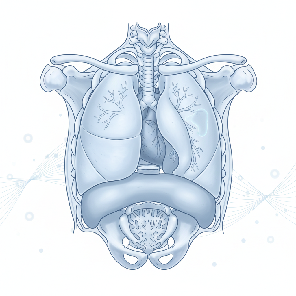
Warning signs that deserve a specialist’s eyes
Many are blamed on weather, age or an old cough. Persistence is what matters — especially when several appear together.
- ①A new cough lasting more than three weeks that refuses to settle.
- ②A long-standing cough changing character — deeper, harsher, or productive differently.
- ③Breathlessness increasing beyond your usual effort level.
- ④Blood-streaked sputum, even in tiny amounts, even once.
- ⑤Chest pain on one side, constant rather than tied to breathing alone.
- ⑥Hoarseness persisting without any throat cause.
- ⑦Pneumonia recurring on the same side — treated each time but always returning to the identical spot.
- ⑧Finger clubbing — fingertips widening and nails curving like watch glasses.
Cancers we treat in this region
From peripheral nodules to central airway tumours and the chest wall itself.
◆Lung Cancer — Keyhole Era
Early lung cancers resected by VATS lobectomy or segmentectomy through keyhole incisions.
◆Central Lung Tumours
Sleeve resections and, when unavoidable, open lobectomy or pneumonectomy with reconstruction.
◆Mediastinal Masses & Thymoma
Tumours between the lungs — including thymectomy for myasthenia-related thymic disease.
◆Pleural Disease
Recurrent effusions managed with pleurodesis so fluid stops stealing breath.
◆Mesothelioma
Pleural malignancy assessed honestly and coordinated with systemic therapy pathways.
◆Chest Wall Tumours
En-bloc resection with skeletal and soft-tissue reconstruction where required.
Operations performed here
- ✦VATS lobectomy / segmentectomyKeyhole removal of a lobe — or less, when anatomy allows — preserving maximum function.
- ✦Open lobectomy & pneumonectomyReserved for cases where open technique genuinely serves safety and completeness.
- ✦Sleeve resectionsTumour removed with airway rejoined end-to-end — curing central cancers while sparing whole lobes.
- ✦Thymectomy & mediastinal mass excisionSafe removal of tumours in the space between the lungs.
- ✦PleurodesisSealing the pleural space so recurring fluid stops compressing the lung.
- ✦Chest wall resection & reconstructionRigorous en-bloc clearance rebuilt for strength and appearance.
From first visit to full recovery
CT & biopsy
The nodule characterised properly before anyone mentions surgery.
Lung function testing
Your reserve measured first — it decides how much lung can safely leave.
Staging & board plan
Nodes mapped; medical and radiation oncology consulted early.
Surgery — VATS-first
Keyhole unless open access truly serves you better.
Early mobilisation
Chest physiotherapy from day one; breathing exercises coached, not rushed.
Surveillance
Scheduled CT reviews catch anything new at its smallest.
Families usually ask…
For most early lung cancers, VATS offers equal cancer outcomes with less pain and faster recovery. But neither approach is universally superior: some central and previously-treated tumours are safer open. The recommendation follows your tumour’s position and your lung reserve — not preference.
Most patients return to normal daily activity — many climb stairs, travel, exercise. The remaining lung expands and its efficiency improves over months. This is exactly why lung-function testing happens before planning: we operate within your reserve, never past it.
Almost always yes — treatment differs completely for infection, scarring or lymphoma versus lung cancer, and biopsy prevents operations that shouldn’t happen. Rarely, a highly suspicious scan pattern justifies going straight to excision; even then it is a considered exception, not a shortcut.
Node-positive disease often still benefits from surgery — frequently combined with chemotherapy before or after. Mediastinoscopy or staging scans tell us which stations are involved, and the multidisciplinary team plans around them. Node status changes the sequence, rarely the commitment.
Any reduction helps wounds heal and lungs clear secretions — even a few weeks makes measurable difference. Support is available whenever you want it, structured and judgment-free. It is your decision; our job is to make the attempt easier, not to lecture.